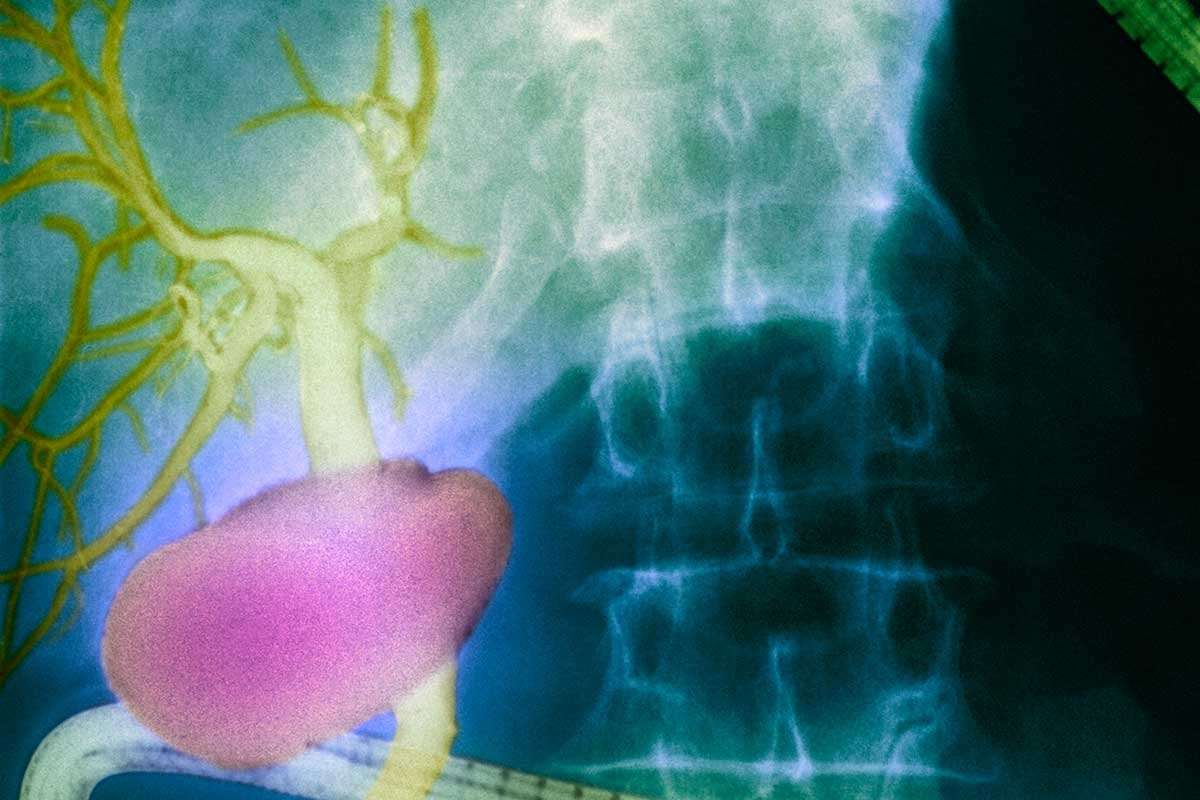
A newly discovered network of fluid-filled channels in the human body may be a previously-unknown organ, and it seems to help transport cancer cells around the body.
This discovery was made by chance, from routine endoscopies – a procedure that involves inserting a thin camera into a person’s gastrointestinal tract. Newer approaches enable doctors to use this procedure to get a microscopic look at the tissue inside a person’s gut at the same time, with some surprising results.
Read more: Cancer algorithm uses game theory to double survival time
One team had expected to find that the bile duct is surrounded by a hard, dense wall of tissue. But instead, they saw weird, unexplained patterns. They took their findings to Neil Theise, a pathologist at New York University School of Medicine.
When Theise used the same endomicroscopy device to look under the skin of his own nose, he saw a similar result. Further investigation of other organs suggested that these patterns are made by a type of fluid moving through channels that are everywhere in the body.
Theise reckons that every tissue in the body may be surrounded by a network of these channels, which essentially form an organ. The team estimate that the organ contains around a fifth of the total fluid volume of the human body. “We think they act as shock absorbers,” says Theise.
This organ was likely never seen before because standard approaches for processing and visualising human tissue causes the channels to drain, and the collagen fibres that give the network its structure to collapse in on themselves. This would have made the channels appear like a hard wall of dense protective tissue, instead of a fluid-filled cushion.
But as well as protecting organs, the network may also aid the spread of cancer. When Theise’s team looked at samples taken from people with invasive cancers, they found evidence that cancer cells that had worked their way out of their original tissues could find their way into these channels, which took them directly to the lymphatic system. “Once they get in, it’s like they’re on a water slide,” says Theise. “We have a new window on the mechanism of tumour spread.”
Theise and his colleagues are now investigating whether analysing the fluid in these newly-discovered channels might lead to earlier diagnosis of cancers. They think the organ might also be involved in other problems, including oedema, a rare liver disease, and other inflammatory disorders.
Journal reference: Scientific Reports, DOI: 10.1038/s41598-018-23062-6
Read more: Bacteria on our bodies may be protecting us from skin cancer; Cancer’s penicillin moment: Drugs that unleash the immune system; Upgraded Pap test detects two extra cancers before they spread; First timeline of a cancer tracks tumours from origin to spread
ChickenInvader42 on March 27th, 2018 at 14:09 UTC »
I think that this discovery only expands our knowledge of the interstitium and it's connection to the lymph nodes, I wouldn't go as far as call it a new organ.
ThomasCro on March 27th, 2018 at 13:03 UTC »
How can it be possible that thousands of autopsies have never revealed this "organ" before?
mvea on March 27th, 2018 at 11:15 UTC »
The post title is a copy and paste from the first paragraph of the linked popular press article here :
Journal Reference:
Structure and Distribution of an Unrecognized Interstitium in Human Tissues
Petros C. Benias, Rebecca G. Wells, Bridget Sackey-Aboagye, Heather Klavan, Jason Reidy, Darren Buonocore, Markus Miranda, Susan Kornacki, Michael Wayne, David L. Carr-Locke & Neil D. Theise
Scientific Reports, volume 8, Article number: 4947 (2018)
doi:10.1038/s41598-018-23062-6
Link: https://www.nature.com/articles/s41598-018-23062-6
Published online: 27 March 2018
Abstract