Male and female adult (2–3 months) mice of C57BL/6J and CD-1 strains were used. Mice were housed in a specific pathogen-free and temperature- and humidity-controlled facility under a light–dark cycle (10 h light and 14 h dark) with food and water ad libitum at the University of Nevada, Reno. Animal use protocol was approved by Institutional Animal Care and Use Committee of the University of Nevada, Reno, and is in accordance with the “Guide for the Care and Use of Experimental Animals” established by National Institutes of Health (1996, revised 2011). All cynomolgus monkeys used were housed at the Blooming Spring Biological Technology Development Co. LTD, in Guangzhou, China, which is fully accredited by the Association for the Assessment and Accreditations of Laboratory Animal Care International. The animal use protocol was approved by the Research Ethics Committee of the Blooming Spring Biological Technology Development Co. LTD. A total of 12 healthy male adult (9–13 years of age with body weight ranging between 4.96 and 11.80 kg) and 6 fertility-proven adult females (9–13 years of age with body weight ranging between 3.89 and 4.00 kg) cynomolgus monkeys were used in this study. The general information of the monkeys used in this study is summarized in Supplementary Table 4.
Triptonide (C 20 H 22 O 6 , MW: 356.39 g/mol) was purchased from the Chengdu Biopurity Phytochemicals (purity > 98%, Lot#: 15033012 and 14081502) and MCE (MedChem Express; purity > 98%, Cas#: 38647-11-9. Lot#:13216). DMSO was purchased from Sigma-Aldrich. In addition to HPLC data provided by the company, the purity of triptonide used was independently validated using NMR elemental analyses in NuMega Resonance Labs, Inc. (San Diego, CA; Supplementary Fig. 15).
For POC efficacy testing on mice, a stock solution of triptonide was prepared at 4 mg/ml in DMSO. Each vial was wrapped with aluminum foil to block light and stored at −20 °C. For gavage feeding, 125 μl of triptonide working solution (5 μl stock solution was added into 120 μl of PBS) containing 20 μg triptonide were administered to a mouse of 25 g (0.8 mg/kg B.W) using a gavage feeding needle (20 G 1–1/2 in., Cadence Science, Japan). Other doses (0.1, 0.2, 0.4, and 1.6 mg/kg BW) were prepared accordingly. Control mice received vehicle (4% DMSO in PBS). After oral administration of triptonide or vehicle, the mice were observed every hour for the first 3 h, followed by daily monitoring thereafter.
In the initial pilot study to identify minimal effective dose and duration, five doses (0.1, 0.2, 0.4, 0.8, and 1.6 mg/kg BW) were tested using 18 mice per dose group. Three mice from each dose group were sacrificed weekly to examine sperm parameters and testicular histology for up to 6 weeks. In the subsequent official POC efficacy testing, 6–20 mice in each test group were treated with triptonide at one of the four doses (0.1, 0.2, 0.4, and 0.8 mg/kg BW) via single daily oral gavage. At the end of the fourth week, all triptonide-treated and control mice were sacrificed and caudal epididymal sperm parameters were measured using a computer-assisted sperm analysis (CASA) system (Sperm Analyzer Mouse Traxx, Hamilton-Thorne). In addition, blood samples were collected and allowed to clot at room temperature for 1.5 h. After disrupting clot adhesion to the tube wall, the blood samples were centrifuged at 2000 × g for 10 min at room temperature. Serum was collected into a polypropylene microcentrifuge tube and stored at −20 °C for hormonal measurements. Hormonal levels were measured at the Ligand Assay and Analysis Core, Center for Research in Reproduction, University of Virginia School of Medicine, Charlottesville, Virginia. The Mouse Pituitary Magnetic Bead Panel Multiplex Kit (Cat.# MPTMAG-49K; Lot#3141797; Millipore Corporation, Billerica, MA) was used to determine the levels of FHS and LH following the manufacturer’s instructions. The reportable range the FSH/LH assays was between 0.48 and 300 ng/ml, and the intra-assay CV was between 0.4 and 2.9% for FSH, and 3 and 7.8% for LH. To measure intratesticular testosterone levels, testicular homogenates were prepared by homogenizing the testis in cold (4 °C) PBS (without detergent or EDTA) on ice for 60 s followed by centrifugation (2000 × g for 10 min at 4 °C) to remove cell debris. The supernatants were then collected into 1.5 ml Eppendorf tubes and stored at −80 °C. Both serum and testicular testosterone levels were assayed using the Testosterone Mouse & Rat ELISA Kit (Cat. #IB79106; Kit Lot# 28K088-2; Immuno-Biological Laboratories, Inc., Minneapolis, MN) following the manufacturer’s instructions. The assay is a solid phase enzyme-linked immunosorbent assay based on the principle of competitive binding. The microwells coated with a monoclonal antibody against a unique antigenic site of testosterone were first incubated with the samples so that testosterone in the samples competes with testosterone-conjugated horseradish peroxidase. After washing to remove the unbound molecules, the solid phase was then incubated with the substrate and the colorimetric reaction was used to quantify the levels of testosterone in the samples. A standard curve was first constructed by plotting OD values against concentrations of the standards, and the concentration of unknown samples were determined using this standard curve. The reportable range of this assay was between 8.3 and 1600 ng/dl, and intra-assay CV was between 0.8 and 3.7%.
Histology of all major organs including heart, liver, spleen, lung, lung, brain, colon, small intestine, testis, and male reproductive tracts (epididymis, seminal vesicle, and prostate) was analyzed in-house. Testes were fixed in Bouin’s fixatives and other organs were fixed in 10% neutral buffered formalin followed by embedding into paraffin. Sections were cut followed stained using hematoxylin and eosin for microscopic evaluation.
To test fertility, after 3–4 weeks of oral administrations of triptonide and vehicle control, two or three females were added to individual cages containing single triptonide-treated or control male mice. The female mice were examined every morning for the presence/absence of vaginal plugs. Once plugs were identified, the female mice were removed and after 7 days, they were transferred to a new cage, and the pregnancies and pups were observed and recorded.
Electron microscopy of mouse seminiferous tubules and sperm
Both scanning and transmission electron microscopic analyses were conducted as described5. Scanning electron microscopy was conducted using stages VII and VIII seminiferous tubules dissected from control and triptonide-treated testes, whereas TEM was performed on sperm collected from the cauda epididymis of control and triptonide-treated male mice.
Mouse ICSI was performed as described5. Cauda epididymal sperm were collected from control and triptonide-treated male mice. Only sperm heads were injected into MII oocytes from donor female mice. To study developmental potential of the preimplantation embryos, the injected oocytes were cultured in vitro. For evaluation of full-term development, the 2-pronuclear embryos were transferred into the recipient females.
Proof-of-concept efficacy testing in cynomolgus monkeys
To identify minimal effective dose and duration, four cynomolgus monkeys were treated with triptonide at four doses (0.05, 0.1, 0.2, 0.8, and 5 mg/kg BW) daily. Based on the weight of each monkey, the total amount of triptonide equivalent to 40 days of oral treatment of 0.1 mg/kg BW was calculated and weighted for making the stock solution in 2 ml DMSO. The working solution was prepared by diluting the stock solution with 18 ml PBS followed by aliquoting into 40 vials (0.5 ml each) for storage at −20 °C. One vial was thawed each day and the triptonide working solution (0.5 ml) was added into the food (steamed bun, bread, apple, banana, etc.) before feeding. The technician observed the feeding to make sure the triptonide-containing food was completely eaten. By the end of each week, semen and blood samples were collected for analyses of sperm parameters and blood chemistry. The pilot test was stopped at week 9 because almost all sperm became deformed with minimal or no forward motility and sperm counts had been drastically reduced. One monkey that received 5 mg/kg BW triptonide daily p.o. displayed signs of side effects starting at week 4 and became worse thereafter; thus, the experiment was stopped at the end of week 5 and that monkey was not used in subsequent POC efficacy testing.
Based on the pilot test, the minimal effective dose was determined to be 0.1 mg/kg (BW). In the official POC efficacy testing, seven male monkeys were treated with single daily doses of triptonide (0.1 mg/kg BW) and three control male monkeys received vehicle. Since we already knew that significant effects on sperm production started at week 4, semen and blood samples were collected weekly starting from week 5. By the end of week 8, triptonide treatment was stopped in three of the seven monkeys, and the other four continued with triptonide treatment until week 126 (~2.4 years). For the monkeys undergoing long-term treatment, semen and blood samples were collected at 11, 14, 16, 18, 23, 48, 74, 100, and 126 weeks for semen and blood chemistry analyses.
To test fertility, two out of the three monkeys with treatment stopped at week 8 were individually housed with two fertility-proven adult female monkeys (one male and one female per cage) for up to 1 year. Both female monkeys became pregnant after 3–4 months and each delivered a full-term baby. Two out of the four male monkeys undergoing long-term treatment were also housed with two fertility-proven adult female monkeys (one male and one female per cage) between weeks 8 and 126. None of the two females became pregnant during the entire treatment. Two of the three controls were housed with two fertility-proven adult female monkeys (one male and one female per cage) between weeks 8 and 100. Both female monkeys became pregnant after 3–4 months, but one ended up with abortion for unknown reasons, but the other delivered a full-term baby.
For semen collection, a rectal probe electrical stimulation method was conducted as previously described45 with minor modifications. Monkeys were anesthetized with ketamine hydrochloride (Lianyungang International Trade Co., Ltd., Lianyungang City, China) at a dose of 10 mg/kg BW through intramuscular injection. The monkeys were then held in a supine position on an operating table and penis was cleaned using absorbent cotton soaked with warmed saline. A lubricated probe was inserted into rectum for ~8–10 cm and positioned with both electrodes oriented in mid-ventral direction. A stimulator with nine-step voltage control (Lane Manufacturing Inc., USA) was used, and each electrical stimulation consisted of pulses of 4–6 s duration with 2–3 s rest in between. Stimulation started at step 1 for five repeats. If no painful response was observed, stimulation was continued with the voltage raised to next step. The stimulation was repeated and gradually intensified until ejaculation occurred. The ejaculates were collected into a tube and incubated in 37 °C water bath for 30 min before analyses. Semen samples were diluted using a sperm culture medium (K-SISM-20/50/10020, Cook Medical) to an appropriate concentration, and an aliquot of 10 μl was added to a pre-warmed (37 °C) Makler counting chamber and sperm counts, total and forward motility were analyzed manually by an experienced evaluator. For motility evaluation, at least 300 sperm were analyzed. For sperm morphology analyses, sperm smear was prepared. After air dry, the slides were fixed in 95% ethanol for 15 min at room temperature, followed by staining using SpermBlue fixative and SpermBlue stain45.
To analyze testicular histology, testicular biopsy was performed under anesthesia (ketamine hydrochloride at 10 mg/kg BW i.m.) and sterile conditions. A small piece of testis (~3 mm × 3 mm × 3 mm) was surgically removed, and immediately fixed in Bouin’s fixative. After dehydration, the samples were embedded into paraffin and sections of 4 μm were then stained using hematoxylin-eosin followed by microscopic evaluation.
Blood oxygen saturation levels were measured using a pulse oximeter (Yuwell, YX303, Yunyue Medical Equipment, Ltd, Jiangsu, China) by clamping the oximeter to the tail of the monkey. Blood assays included counts of the red blood cells, lymphocytes, mean corpuscular volume, and levels of prothrombin, hemoglobin, and glucose. The liver panel contained the following: albumin-globulin, albumin, alkaline phosphatase, alanine aminotransferase, aspartate aminotransferase, carbon dioxide, gamma-glutamyl transferase, and total protein. The kidney panel consisted of creatinine, urine albumin, and urea. All the assays were conducted in the Central Clinical Laboratory of the Hospital affiliated to the Institute of Family Panning of Guangdong Province (Guangzhou, China) using the reagents, equipment, and methodologies for human blood lab work. In brief, blood cell types and biochemistry were analyzed using a hematology analyzer (Sysmex XN-1000, Japan). FSH, LH, and T levels were measured using fluorescent immunoassays on an immunology analyzer (Roche Cobas E602, Switzerland). Liver and kidney panels were analyzed using a clinical chemistry analyzer (Olympus, AU400, Japan). Assays were conducted using reagents and protocols provided by the manufactures of the equipment.
Determination of 50% of effective concentration
EC 50 represents the dose at which a compound produces half of its maximal effect46. To determine the EC 50 of triptonide, a series of dose–response data were collected using adult male mice treated with various doses of triptonide in three independent experiments. Sperm motility was evaluated using CASA (Hamilton Throne). To determine male fertility, two to three fertility-proven females were added to the cages containing a single triptonide-treated or control male mouse at the end of the fourth weeks of treatment. Vaginal plugs were examined early every morning to verify mating, and pregnancy became visible by day 10 after mating. Later, the number of pups borne was documented (Supplemental Table S3). EC 50 was calculated based on a basic rectilinear equation: E = mx + E 0 , where E represents the triptonide effect, m stands for the slope (\(\frac{{{\mathbf{E}}_0 - {\mathbf{E}}_{0.1}}}{{0 - 0.1}}\)), and x for triptonide concentration (C). Since E = \(\frac{{{\mathbf{E}}_0 - {\mathbf{E}}_{0.1}}}{{0 - 0.1}}\) C + E 0, when x = 0, the E equals E 0 (E = E 0 ). Because E = \(\frac{{{\mathbf{E}}_0 - {\mathbf{E}}_{0.1}}}{{0 - 0.1}}\) C + E 0 , where E 0.1 equals E, when C = 0.1. When E = \(\frac{{{\mathbf{E}}_0}}{2}\), the C = C 0.5. Since E = \(\left( {\frac{{{\mathbf{E}}_0 - {\mathbf{E}}_{0.1}}}{{0 - 0.1}}} \right)\;{\mathbf{C}} + {\mathbf{E}}_0\); \(\frac{{{\mathbf{E}}_0}}{2} = \frac{{{\mathbf{E}}_0 - {\mathbf{E}}_{0.1}}}{{0 - 0.1}}\) C 0.5 + E 0 , and \(\frac{{{\mathbf{E}}_0}}{2}\)= M C 0.5 + E 0. Thus, EC 0.5 = \(\frac{{\frac{{{\mathbf{E}}_0}}{2} - {\mathbf{E}}_0}}{{\mathbf{M}}} = \frac{{ - \frac{{{\mathbf{E}}_0}}{2}}}{{\mathbf{M}}} = - \frac{{{\mathbf{E}}_0}}{{2{\mathbf{M}}}}.\) Specifically, we utilized E 0 (sperm motility or male fertility of control male mice) through its slope \(\left( {\frac{{{\mathbf{E}}_0 - {\mathbf{E}}_{0.1}}}{{0 - 0.1}}} \right),\)and the equation (C 0.5 = −E 0 /2m), to acquire the EC 50 .
Testes were lysed in Pierce IP lysis buffer (Thermo Scientific, no.87787, 25 mM Tris HCl, pH 7.4, 150 mM NaCl, 1% NP-40, 1 mM EDTA, and 5% glycerol) containing protease inhibitors (Roche, mini-complete, no EDTA) and homogenized. The lysates were vortexed and spun at 17,000 × g, 20 °C for 10 min, and the supernatants were collected. The protein concentrations were determined using Pierce BCA Protein Assay Kit (Thermo Scientific). The proteins from each sample were loaded onto one MiniProtean TGX 4–15% or 4–20% polyacrylamide gel (Bio-Rad) followed by electrophoresis at 200 V for 40 min. The gel was electroblotted onto nitrocellulose membrane at 100 V for 1 h in a cold room. After blocking with SuperBlock T20 (PBS) Blocking Buffer (Thermo Fisher Scientific) and the membrane was subjected to reactions with primary antibodies on a rocking platform in a cool room overnight. After washing, the membrane was incubated with goat anti-mouse IgG HRP or goat anti-rabbit IgG HRP for 1 h. The specific proteins were visualized using the Advanced Bright Enhanced Chemiluminescence kit. Imaging and qualification were performed using a ChemiDoc Imager Detector (Bio-Rad). The primary antibodies used included mouse monoclonal anti-γH2AX (phospS139, Abcam, Cat#: 2635), rabbit polyclonal anti-β-actin (Abcam, Cat#: 8227), rabbit polyclonal keratin 5 (K5; BioLegend, Cat#:905501), and mouse monoclonal anti-junction plakoglobin/gamma catenine (Life Technologies, Cat#:13-8500). Goat anti-rabbit (SouthernBiotech, Cat#: 4030-05) and goat anti-mouse IgG (H + L; SouthernBiotech, Cat#: 1036-05) HRP-conjugated secondary antibodies were purchased from SouthernBiotech. Original uncropped western blot scans or films can be found in the Source data file.
Drug affinity responsive target stability assay
Four testes were removed from adult male C57/BL/6J mice. Each testis was dissected into several pieces and put into a 1.5 ml tube containing 450 μl M-PER solution (Thermo Fisher Scientific) with protease inhibitors (Roche, complete mini, EDTA-free) and phosphatase inhibitors (Abcam, Phosphatase inhibitor cocktail I). The mixture was homogenized with 20 strokes of a plastic pestle and then homogenized with a Benchmark D1000 homogenizer at a setting of two for 15 s. All four testis samples sat on ice for a half hour and were then centrifuged at 17,200 × g for 10 min. The four supernatants were pooled in a 15 ml centrifuge tube. To the tube was added 200 μl 10× TNC (500 mM Tris HCl, 500 mM NaCl, 100 mM CaCl 2 , pH 8.0). The homogenate was assayed for protein concentration with a BCA assay and was diluted to 5 mg/ml. Homogenate (297 μl) was aliquoted into two tubes. To the tubes were added either 3 μl 16 mg/ml triptonide in DMSO or DMSO alone. The tubes were mixed well and left at room temperature for 1 h. Meanwhile, a solution of 10 mg/ml Pronase was diluted as follows: 1:100, 1:300, 1:1000, 1:3000, and 1:10,000. When Thermolysin was used in place of Pronase, a 10 mg/ml solution of Thermolysin was diluted to 2, 0.4, 0.08, and 0.016 mg/ml. The triptonide-treated or control homogenate (50 μl) was incubated with each of trypsin dilution for 15 min. The reactions were then stopped by the addition of 5 μl 0.5 M EDTA. Each sample was then run on Bio-Rad mini-Protein TGX 4-20% polyacrylamide gels at 200 V. Gels were stained with Bio-Safe Coomassie (Bio-Rad).
An in vivo variation of the DARTS experiment was carried out using testis homogenates from mice treated with 0.8 mg/kg BW triptonide daily (p.o.) for 4 weeks. Protein was precipitated from these extracts with four volumes of ice-cold acetone. After overnight incubation at −20 °C, the samples were centrifuged at 16,000 × g, 4 °C, for 10 min. The pellet was washed three times with ice-cold acetone/water (4:1). The protein pellet was dried, dissolved, and digested for various lengths of time using the Flash Digest system from Perfinity Biosciences. The digest reaction was stopped after 0, 5, 15, and 75 min of incubation by removal of aliquots and addition into Laemmli buffer with β-mercaptoethanol. The aliquots were run on polyacrylamide gels followed by visualization of protein using staining. Bands of interest were cut out and submitted for MS analyses. Original gel images can be found in the source data file.
GlycoLink Micro Immobilization Kit was purchased from Thermo Fisher Scientific. GlycoLink beads contain a linker with a terminal hydrazide group which reacts with aldehydes and ketones. The coupling of triptonide to the beads was attempted in two ways. The GlycoLink kit comes with an acidic coupling buffer for coupling to aldehydes and ketones. An additional basic buffer was purchased which might couple via the epoxide groups of triptonide. Coupling was done by incubating beads with a saturated solution of triptonide in DMSO (40 mg/ml), or with DMSO alone for controls, in acidic and basic buffer. Triptonide was covalently attached to the beads with aniline according to the manufacturer’s instructions. Testes from one sexually mature male mouse were homogenized in M-PER buffer (Thermo Fisher Scientific). The proteins in the homogenate were precipitated with four volumes of ice-cold acetone. After sitting for 3.5 h at −20 °C, the protein pellet was centrifuged at 16,000 × g, 4 °C, for 10 min. The pellet was washed three times with ice-cold 4:1 acetone/water. The final pellet was dried and dissolved in 2.5 ml Dulbecco’s PBS. An aliquot (300 μl) was applied to each of the four types of beads and reacted with the beads as per instructions. After washing of the beads, proteins on the beads were eluted sequentially with the following solutions: a saturated solution of triptonide in DMSO in PBS, 0.1% formic acid/30% acetonitrile, and 0.2 M glycine, pH 2.6. The eluates were dried in a vacuum concentrator to ~60–70 μl. The eluates were then desalted with Zeba Spin Desalting Kit (Thermo Fisher Scientific, 7 K MWCO) and buffer exchanged into 0.1% formic acid/30% acetonitrile. The eluates were again dried to a volume of 35–40 μl. The eluates were then applied to a Bio-Rad mini-Protean TGX 4-20% polyacrylamide gels and run at 200 V. Gels were stained with Sypro Ruby. Bands of interest were cut out and submitted for MS analyses. Original gel images can be found in the Source data file.
Immunoprecipitation-based identification of proteins interreacting with the N-terminus of SPEM1
A peptide composed of the N-terminal 28 amino acids of murine SPEM1 was synthesized by BioLegend (San Diego, CA). A biotin was attached to the C-terminal amino acid of the peptide. Streptavidin magnetic beads were obtained from GenScript. Beads were incubated with either 0.3 mg of peptide in PBS or with PBS alone. The solutions were incubated with the beads for 1 h and then washed with PBS. A homogenate of testis from a sexually mature male mouse was prepared in PBS with protease inhibitors (Roche, complete mini, EDTA-free). The two types of beads were incubated with 120 μl of the testis homogenate (0.34 mg protein) for 1 h. After washing of the beads, proteins on the beads were eluted sequentially with a saturated solution of triptonide in DMSO in PBS, 0.1% formic acid/30% acetonitrile, and 0.2 M glycine, pH 2.6. The eluates were dried in a vacuum concentrator to ~60–70 μl. The eluates were then desalted with Zeba Spin Desalting Kit (Thermo Fisher Scientific, 7 K MWCO) and buffer exchanged into 0.1% formic acid/30% acetonitrile. The eluates were again dried to a volume of 35–40 μl. Half of each eluate was diluted with Laemmli buffer with β-mercaptoethanol and applied to a Bio-Rad mini-Protean TGX 4–20% polyacrylamide gels and run at 200 V. Gels were stained with Sypro Ruby. The remaining halves of the eluates were submitted for MS analysis.
Proteins in the excised electrophoresis bands were reduced and alkylated using 10 mM dithiothreitol and 100 mM iodoacetamide. Proteins in solutions were denatured with acetonitrile, then reduced and alkylated with 6.7 mM DTT and 13.8 mM iodoacetamide. The proteins were then incubated with sequencing grade modified porcine trypsin (Promega, Fitchburg, WI) in 25 mM ammonium bicarbonate overnight at 37 °C.
Peptide mixtures were separated using an UltiMate 3000 RSL Cnano system (Thermo Scientific, San Jose, CA) on a self-packed UChrom C18 column (100 μm × 35 cm). Elution was performed using a 90 min gradient of solvent B from 2–27% (solvent A 0.1% formic acid, and solvent B acetonitrile, 0.1% formic acid) at 50 °C using a digital Pico View nanospray source (New Objectives, Woburn, MA) that was modified with a custom-built column heater and an ABIRD background suppressor (ESI Source Solutions, Woburn, MA). Briefly, the self-packed column tapered tip was pulled with a laser micropipette puller P-2000 (Sutter Instrument Co, Novato, CA) to an approximate id of 10 µm. The column was packed with 1–2 cm of 5 µm Magic C18 followed by 35 cm of 1.8 µm UChrom C18 (120 A) at 9000 p.s.i., using a nano LC column packing kit (nanoLCMS, Golg River, CA).
Mass spectral analysis was performed using an Orbitrap Fusion mass spectrometer (Thermo Scientific. San Jose, CA). Proteomic analysis was performed using a “Universal” data-dependent method (Eliuk et al. 2014 Thermo Application note). The MS1 precursor selection range was from 400 to 1500 m/z at a resolution of 120 K and intensity threshold of 4.0 × 10⁵. Quadrupole isolation at 0.7T h for MS2 analysis using CID fragmentation in the linear ion trap with a collision energy of 35%. The automatic gain control was set to 1.0 × 10⁷ with a maximum injection time of 250 ms.The instrument was set in a top speed data-dependent mode with a most intense precursor priority. Dynamic exclusion was set to an exclusion duration of 60 s with a 10 ppm tolerance.
Tandem mass spectra were extracted and charge state deconvoluted by Proteome Discover version 2.1. All MS/MS samples were analyzed using Sequest (Thermo Fisher Scientific, San Jose, CA, USA; version 2.0.0.802). Sequest was set up to search a custom C-term fasta, assuming the digestion enzyme trypsin and max number of missed cleavages get to 2. Sequest was searched with a fragment ion mass tolerance of 0.60 Da and a parent ion tolerance 10.0 PPM. Variable modifications included carbamidomethyl of cysteine, oxidation of methionine, deamidation, and acetylation of the N-terminus.
Scaffold (version Scaffold 4.8.2, Proteome Software Inc., Portland, OR) was used to validate MS/MS-based peptide and protein identification. Peptide identifications were accepted if they could be established at >80% probability by the Peptide Prophet algorithm47 with Scaffold delta-mass correction. Protein identifications were accepted if they could be established at greater 99.0% probability and contained at least two identified peptides. Protein probabilities were assigned by the Peptide Prophet algorithm48. Proteins that contained similar peptides and could not be differentiated based on MS/MS analysis alone were grouped to satisfy the principles parsimony.
Graph Pad Prism 7 software (La Jolla, CA, USA) was used for statistical analyses. Data are presented as mean ± SEM unless stated otherwise. Differences in measurements were compared by Kolmogorov–Smirnov t test or one-way analyses of variance (ANOVA) for two groups, and p < 0.05 was considered statistically significant. Two-way ANOVA with Bonferroni multiple comparison test were used to compare differences between groups and timepoints, and adjusted p < 0.05 was considered to be statistically significant.
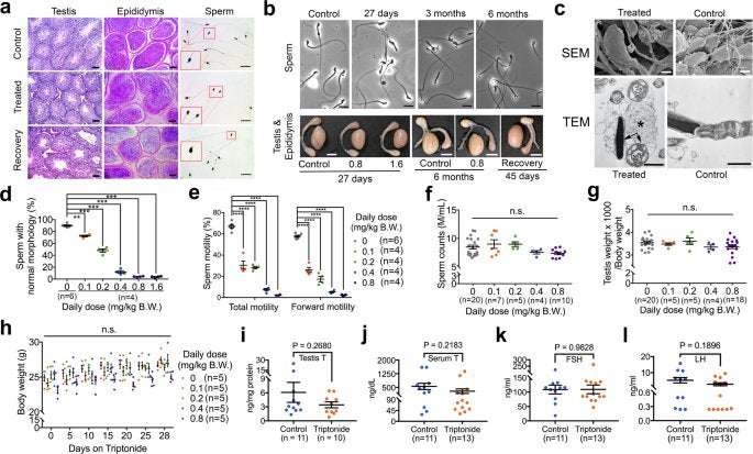
Gross morphology and histology of both the testes and cauda epididymal sperm were routinely examined in both pilot and POC efficacy testing experiments using C57Bl/6J (approximately ten independent experiments expanding 7 years) and CD-1 (two independent experiments in two years) male mice, and results similar to the representative images shown in Figs. 1a, b and Supplementary Fig. 2 were obtained consistently. Both TEM and SEM were conducted once using three mice from three separate experiments, and the data similar to the representative images shown in Fig. 1c were obtained consistently. In the POC efficacy testing on monkeys, ejaculated sperm were collected from all monkeys once every 1–3 weeks, and testicular biopsy was conducted on six treated and three control monkeys at different timepoints. Both sperm morphology and testicular histology varied to some degree, but were generally consistent to the representative images shown in Fig. 2a,e, as well as Supplementary Fig. 4d–i. Pathology examinations at both gross and histological levels were conducted on six treated and three control male C57Bl/6J mice from three independent experiments, and no major pathology was detected and the histology of major vital organs of these mice was similar to the micrographs shown in Supplementary Fig. 3. DARTs were performed twice with similar results, and Fig. 5b is one the two gels obtained and subjected to MS-based protein identification. GlycoLink beads-based affinity purification was repeated four times using samples from four male C57Bl/6J male mice from two independent experiments, and results similar to Fig. 5d–f were obtained. Immunofluorescent and western blot analyses of gamma-H2Ax were carried out three times, using three control and six treated male mice from two independent experiments, and results similar to those shown in Supplementary Fig. 14 were obtained. Efficacy testing using chemically synthesized triptonide was performed twice, using four mice per group per timepoint, and both testicular histology and sperm morphology were similar to those shown in Supplementary Fig. 16.
Further information on research design is available in the Nature Research Reporting Summary linked to this article.
AlexReinkingYale on February 26th, 2021 at 14:08 UTC »
What ever happened to RISUG or Vasalgel or whatever it was called?
sodium_dodecyl on February 26th, 2021 at 13:20 UTC »
Very cool, thanks for sharing. Glad they tested for DSB formation -- always a concern when there's otherwise difficult to explain fertility.
For anyone who doesn't know, Nature makes the peer review file public as well (found here). It can sometimes be an interesting read.
mvea on February 26th, 2021 at 12:17 UTC »
I’ve linked to the open access full-text source journal article in the post. The citation of the article is here:
Triptonide is a reversible non-hormonal male contraceptive agent in mice and non-human primates.
Chang, Z., Qin, W., Zheng, H. et al.
Nat Commun 12, 1253 (2021).
Published 23 February 2021
DOI: https://doi.org/10.1038/s41467-021-21517-5
Abstract
There are no non-hormonal male contraceptives currently on the market despite decades of efforts toward the development of “male pills”. Here, we report that triptonide, a natural compound purified from the Chinese herb Tripterygium Wilfordii Hook F displays reversible male contraceptive effects in both mice and monkeys. Single daily oral doses of triptonide induces deformed sperm with minimal or no forward motility (close to 100% penetrance) and consequently male infertility in 3–4 and 5–6 weeks in mice and cynomolgus monkeys, respectively. Male fertility is regained in ~4–6 weeks after cessation of triptonide intake in both species. Either short- or long-term triptonide treatment causes no discernable systematic toxic side effects based on histological examination of vital organs in mice and hematological and serum biochemical analyses in monkeys. Triptonide appears to target junction plakoglobin and disrupts its interactions with SPEM1 during spermiogenesis. Our data further prove that targeting late spermiogenesis represents an effective strategy for developing non-hormonal male contraceptives.